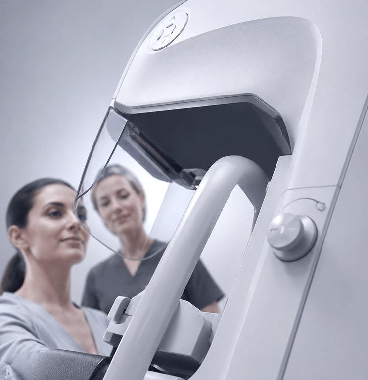
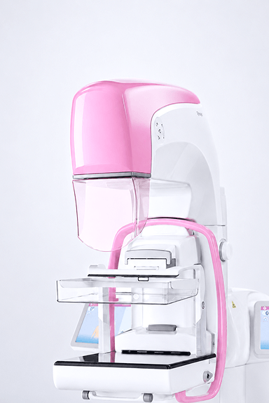
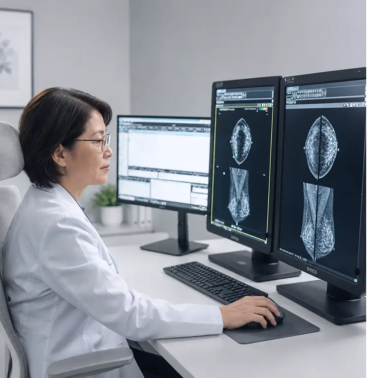
Advanced 2D and 3D mammography technologies providing high-resolution breast imaging for screening, diagnosis, and breast cancer detection. Tomosynthesis imaging supports improved lesion visualization and diagnostic confidence through detailed layered breast imaging.
2D and 3D mammography systems support high-resolution breast imaging with detailed visualization for screening and diagnostic workflows. These technologies help medical teams capture clear, consistent images while supporting accurate evaluation across routine and advanced breast care applications.
With tomosynthesis imaging, clinicians can review layered breast images for improved visibility of structures and potential abnormalities. This advanced approach supports greater diagnostic confidence, helping healthcare providers deliver more precise, efficient, and patient-focused breast imaging services.
01
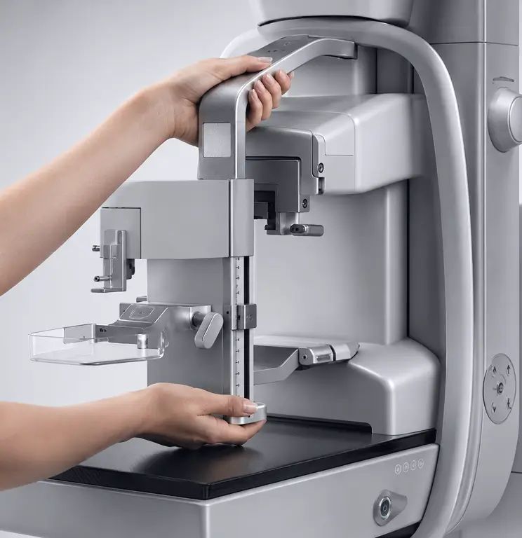
Planmed Clarity™ 2D is a comprehensive digital mammography system. The durable unit can be used for screening, diagnostic examinations, and stereotactic biopsies. Planmed Clarity 2D system comprises all the features of the Planmed Clarity™ product family.
02
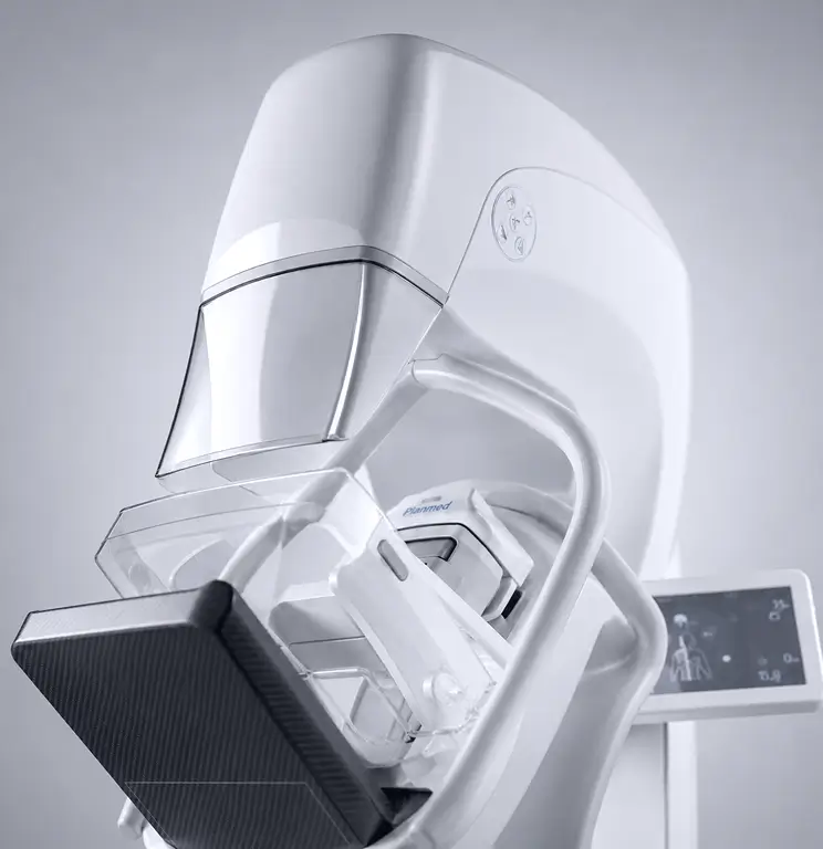
The outstanding image quality and high performance makes Planmed Clarity™ 3D one of the most advanced mammography systems in the market. The durable and versatile system can be used for 3D imaging, 2D screening, diagnostic examinations, and stereotactic biopsies. Planmed Clarity 3D is the complete mammography system comprising all the features of the Planmed Clarity™ product family.
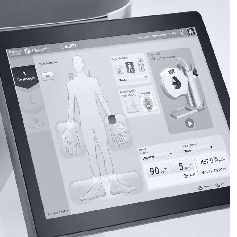
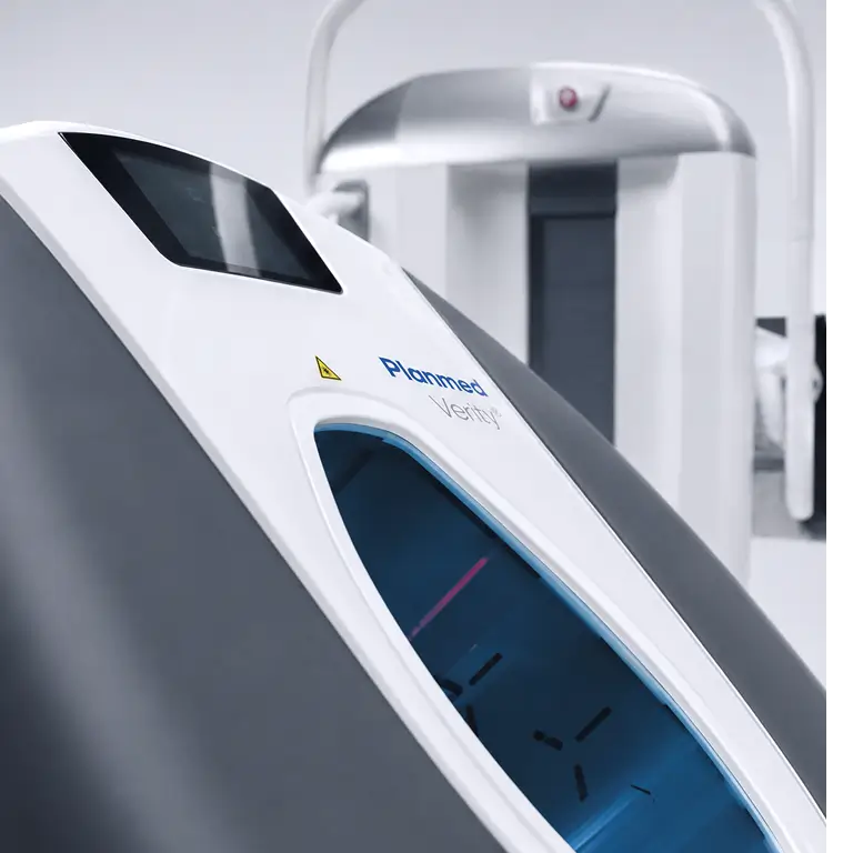
Planmed Verity® CBCT provides high-resolution, low-dose 3D imaging for orthopedic and extremity applications, including weight-bearing imaging under natural load. Designed to support improved fracture diagnostics and treatment planning, the system delivers detailed point-of-care imaging for musculoskeletal, maxillofacial, and head and neck applications.
Cone Beam CT technology supports detailed 3D imaging for orthopedic, extremity, and point-of-care applications. With high-resolution visualization and low-dose performance, it helps clinicians assess complex anatomy with greater clarity while supporting faster, more informed diagnostic workflows.
Designed for musculoskeletal, maxillofacial, and head and neck imaging, Cone Beam CT provides flexible scanning capabilities, including weight-bearing imaging under natural load. This allows medical teams to evaluate fractures, alignment, and treatment planning with a more accurate view of real-life patient positioning.
01
A 3D weight-bearing image of a knee, ankle, foot and toes under natural load can reveal problems otherwise non-discernible. The versatile patient positioning options combined with advanced imaging algorithms allow effortless imaging.
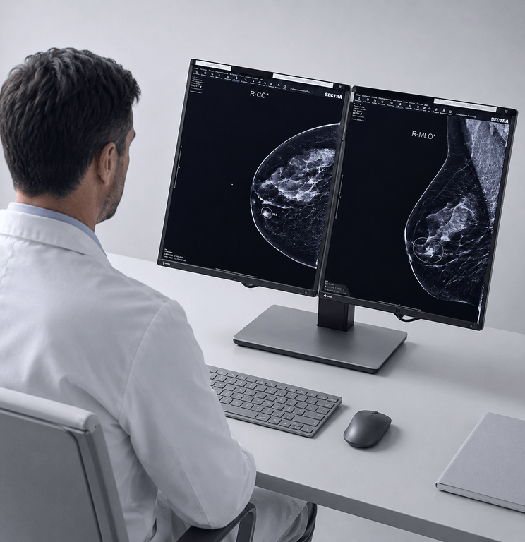
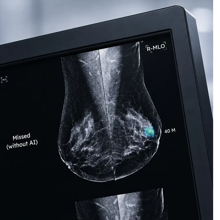
Lunit AI for Mammography provides advanced image analysis tools designed to assist radiologists during breast cancer screening. By supporting lesion detection, improving diagnostic accuracy, and streamlining workflow efficiency, AI-assisted mammography technologies help enable earlier detection and more confident clinical decision-making.
AI for mammography supports radiologists by analyzing breast images with advanced detection tools that help identify suspicious findings more efficiently. By adding an intelligent layer to the screening process, it helps improve consistency, support diagnostic accuracy, and assist clinical teams during high-volume breast imaging workflows.
Designed to complement radiologist expertise, AI-assisted mammography helps streamline image review, prioritize cases, and support earlier breast cancer detection. This technology enhances clinical confidence by providing additional insights that can improve decision-making and create a more efficient, patient-focused screening experience.
01
A globally trusted AI software that delivers smarter, earlier breast cancer detection.
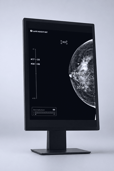
02
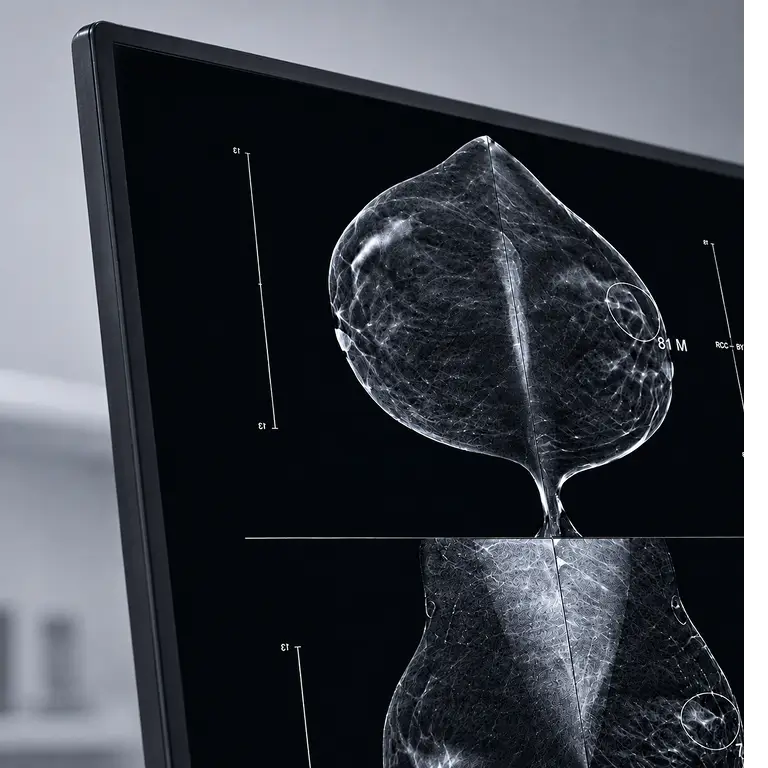
Analyze multi-slice exams with AI that highlights suspicious findings, speeds up review, and reduces reading fatigue.
Connect with our team to explore the right solutions for your clinical environment and operational needs.